
Fluorophores have long enabled biologists to localize and quantify features present in a given sample. Proteins and dyes capable of fluorescence can be used to stain targets in a sample or introduced into your sample’s genome and colocalized with a target protein as a marker of that protein’s expression. Targets that are selected for analysis with fluorescent molecules are not always perfectly flat and 3d information can be useful in determining the function of the target that a fluorophore labels. Two-photon fluorescent microscopy takes advantage of a reduction in scattering of light in the infrared range in tissue to view fluorophores beneath the surface of a sample.
Electrons are capable of moving to different energy levels after receiving a specific amount of energy. When relaxing back into their resting energy levels they sometime emit a photon in the visible light range. Fluorophores are molecules that are capable of emitting a photon after receiving a photon of greater energy than the photon they emit. The energy a photon can be calculated by multiplying Plank’s constant by the speed of light and dividing by the wavelength:
\[E = \frac{hc}{\lambda}\]
There are cases where a fluorophore can emit a photon without having come into contact with a single photon at the correct excitation wavelength. In Dirac’s 1927 paper The Quantum Theory of Dispersion he noted that a few terms of one of his equations would correspond to two light quanta being simultaneously absorbed, but also noted that they would not contribute to the overall result as this event is highly unlikely [1]. Maria Goppert-Mayer described this two-photon excitation fully in her 1931 dissertation [2]. When absorbing two photons simultaneously each photon would need to have half the energy for excitation, or twice the wavelength by the above equation.
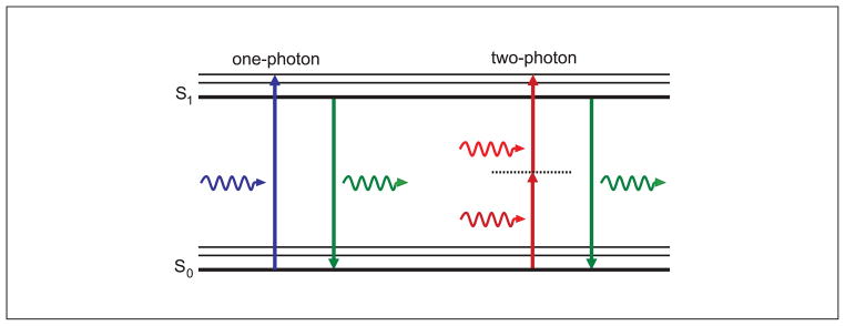
Figure 1: From Two-Photon Excitation Microscopy for the Study of Living Cells and Tissues [3].
In order for two photon absorption to take place, the fluorophore and 2 photons have to be in the same place at the same time, much less than 1 femtosecond (10e-15). In order to make this event of two photons and a fluorophore meeting in such a short time possible, Winfreid Denk implemented high power femtosecond pulsed lasers [4]. These lasers release large quantities of photons in short bursts of about 100 femtoseconds and are focused on a small region of interest [4]. When the light emitted by the laser has twice the wavelength generally required for fluorescence, two-photon fluorescence is possible. Denk coupled this two-photon excitation with laser scanning microscopy where an entire image is acquired by excitation at small regions throughout the target.
The main benefit of this method is that it allows for longer wavelengths to be used to excite the fluorophores, notably in the near infrared range. Wavelengths in this part of the spectrum do not scatter in tissue as much as wavelengths in the visible range do. This allows for two-photon microscopy to image beneath the surface up to a few millimetres beneath the surface.
More information on two-photon microscopy can be found at the following links:
https://www.spiedigitallibrary.org/journals/journal-of-biomedical-optics/volume-25/issue-01/014511/Multiphoton-microscopy--a-personal-historical-review-with-some-future/10.1117/1.JBO.25.1.014511.full#r1
https://www.microscopyu.com/techniques/multi-photon/multiphoton-microscopy

Author